To accurately evaluate oral and dental health, a visual examination alone is often not sufficient. One of the most commonly used imaging methods in dentistry, panoramic dental X-ray, provides a detailed view of the jaw and teeth in a single frame, making it crucial for both diagnosis and treatment planning. In this article, you can find detailed answers to the most frequently asked questions, including what a panoramic X-ray is, how it is taken, which problems it detects, who it is applied to, whether it has any risks, its safety for pregnant women, use in children, and costs.
What is a Panoramic Dental X-Ray?
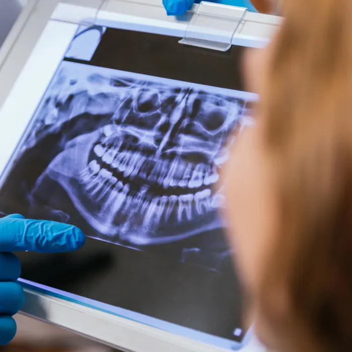
A panoramic dental X-ray is a special dental imaging method that shows all teeth in the upper and lower jaw, the jawbone, jaw joints (TMJ), sinus areas, and surrounding anatomical structures in a single wide image.
- It is a 2D image.
- It takes approximately 10–15 seconds.
- It is completely painless and quick for the patient.
- It provides the dentist with a “large map” of the oral cavity.
Therefore, it is the most commonly preferred X-ray type for initial examination, treatment planning, general oral screening, and detecting hidden problems.
What Does a Panoramic X-Ray Show?
With a panoramic X-ray, the dentist can see many details inside the mouth:
1. Cavities and Tooth Structure
- Interproximal cavities that are not normally visible
- Losses in tooth enamel
- Root structure deformities
2. Gum Diseases and Bone Levels
- Bone loss due to periodontitis
- Inflammation beneath the gums
- Tartar deposits on the root surface
3. Impacted Teeth
- Impacted wisdom teeth (third molars)
- Teeth growing in misaligned positions
- Teeth exerting pressure on the jaw
4. Cysts, Tumors, and Lesions
- Cyst formations
- Tumor-like structures in the bone
- Pathologies spreading to the sinus area
5. Root Tip Infections
- Granulomas
- Abscess formation
- Inflammation around the root
6. Jaw Structure and Joint Problems
- Jaw joint (TMJ) structure
- Bone fractures
- Changes due to trauma
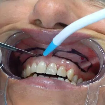
How is a Panoramic X-Ray Taken?
The procedure is quite simple:
- The patient stands in front of the device.
- The jaw, forehead, and cheeks are lightly stabilized with an apparatus.
- Metal accessories are removed.
- The device rotates 360° around the patient.
- The process is completed in 10–15 seconds.
Is the radiation dose high?
The radiation dose of a panoramic X-ray is low, and in modern devices, it is minimized. It is comparable to radiation sources encountered in daily life.
When is a Panoramic X-Ray Taken?
1. General Screening During Initial Examination
The most basic imaging method for the dentist to quickly assess the oral cavity.
2. Detecting Tooth Cavities
It can reveal deep cavities not visible to the eye.
3. Gum Diseases
Important for periodontitis diagnosis as it shows bone loss.
4. Orthodontics (Before Braces)
Provides detailed views of tooth alignment, jawbone structure, and impacted teeth.
5. Before Implant Treatment
- Bone thickness
- Bone height
- Condition of the sinus cavities
Essential data for implant planning is obtained.
6. Before Impacted Tooth Surgery
Provides guidance, especially for wisdom tooth surgery.
7. Jaw Pain and TMJ Problems
Gives information about the joint structure.
Advantages of Panoramic X-Ray
- Entire mouth and jaw in a single image
- Quick and painless
- Low radiation
- Detects hidden dental problems
- Provides accurate planning for implants, orthodontics, and surgery
- Assesses general jaw condition
Is a Panoramic X-Ray Harmful?
The radiation dose in modern digital devices is very low and is lower than many sources of daily radiation.
1 panoramic X-ray ≈ 1–2 days of natural radiation. It is generally not performed during pregnancy unless necessary.
Panoramic X-Ray and Implant Treatment
In implant treatment, the panoramic X-ray is the first step imaging. However, for more detailed information, 3D CBCT (cone beam CT) is often required. The panoramic X-ray provides:
- General idea of bone density
- Sinus height
- Impacted roots
- Bone levels
- Condition of neighboring teeth
It is therefore an integral part of implant planning.
Frequently Asked Questions
Does a panoramic X-ray show all dental problems?
A panoramic X-ray provides a wide view but does not show all details. It offers an initial evaluation of teeth, jawbones, and sinuses, but cannot show micro-level details.
Problems that can be seen on a panoramic X-ray:
- Most cavities
- Root tip infections
- Cyst or tumor-like formations
- Position of wisdom teeth
- Bone loss due to periodontitis
- Jaw structure and symmetry issues
- Impacted or missing teeth
- Trauma-related fractures
- Relationship between sinus floor and tooth roots
Situations where a panoramic X-ray may be insufficient:
- Very small or early-stage cavities
- Root canal details (number, curvature, narrowing)
- Root cracks
- Very small lesions around a tooth
- Millimetric bone measurements needed for implants
In such cases, the dentist may request a periapical X-ray or 3D dental tomography (CBCT).
Is the radiation from a panoramic X-ray harmful?
Modern digital panoramic X-ray devices emit very low radiation, even less than the natural radiation encountered in daily life.
Comparison:
- 1 panoramic X-ray ≈ 1–2 days of natural radiation
- A short airplane flight ≈ 1 panoramic X-ray
- 1 chest X-ray ≈ 4–5 panoramic X-rays
Therefore, a single panoramic X-ray is generally safe for health.
Why it is safe:
- The device emits local, low-dose radiation as it rotates around the patient.
- Radiation levels are limited by international standards.
- Digital sensors use 3–5 times less dose than old analog films.
- Lead aprons and protective devices are used in clinics.
When caution is needed:
- Pregnancy (especially first trimester)
- Frequent and unnecessary X-rays
- Individuals with thyroid sensitivity
- Child patients (lower dose should be used)
Is a panoramic X-ray sufficient before implant treatment?
No. A panoramic X-ray is sufficient for the first step before implants, but precise planning requires 3D CBCT (cone beam CT).
What does a panoramic X-ray provide for implants?
- General idea of bone height
- Sinus cavity position
- Root structure
- Anatomical boundaries
- Overall jaw shape
- Presence of impacted roots or teeth
However, these are approximate values and not cross-sectional images.
Why is CBCT necessary?
3D tomography:
- Measures bone thickness to the millimeter
- Shows nerve canal distance exactly
- Displays sinus membrane in 3D
- Assesses bone density
- Allows implant angle simulation
Panoramic X-ray “provides an image”, CBCT “plans the surgery”.
Do I need a panoramic X-ray? Is it safe?
During pregnancy, X-rays, especially in the first trimester (0–12 weeks), should be avoided if possible. This period is critical for organ development, and even very low doses should be carefully considered.
Basic rules for X-rays during pregnancy:
Not mandatory: Do not take. Routine checks are strictly not recommended.
If necessary, protection must be ensured:
- Lead apron
- Thyroid protector
- Low-dose mode
Most dental procedures can be postponed to the 2nd trimester of pregnancy.
When can it still be taken?
- Severe abscess
- Suspected cyst
- Fracture or trauma
- Dental infection of vital importance
- Rapidly spreading facial infection
In such cases, the X-ray is necessary for the mother’s health and can be performed under the dentist’s evaluation.


